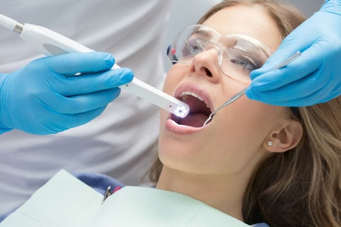
Have you ever found yourself discussing with coworkers about the patient in your schedule who is non-compliant with their treatment plan? The patient has had treatment recommended countless times, only to have them walk out with it unscheduled or has ended up canceling the appointment for said treatment. Working in the dental field, we hear all too often from patients, “The tooth is fine because it is not bothersome and doesn’t hurt,” and how the doctor must be trying to buy a new (insert word here). The list goes on and on. The single best tool for non-compliance? Enter the intraoral camera.
I have always had the best patient compliance when using intraoral cameras. What better way to show patients what is really going on in their mouth? The first time a patient sees their teeth blown up on a screen with a clear color photo, most the time the reaction is one of surprise. “I can’t believe my tooth looks so bad!” Or, “Wow, that is gross.” I’ve even heard, “That’s my mouth?”
Showing the patient leaking or broken restorations and fracture lines in a large view allows the patient to truly understand what is going on in their mouth. Intraoral cameras can be used as a tracking tool as well. Noticing changes in tooth alignment, re-evaluating lesions, calculus buildup, inflamed tissues, or lesions, are just examples of what can be monitored with an intraoral camera.
If your practice does not have an intraoral camera, or it just sits unused, you are missing out!
Most of us are visual learners. It can be hard to comprehend what is being said or explained without seeing a clear picture of exactly what is going on. Our dental patients are no different. Telling the patient about the fracture line on a tooth versus showing them a photo of the area can be a game changer for the patient.
I have had access to an intraoral camera at various points in my career. They would be randomly used to capture an image of a broken tooth, abscesses, or heavy calculus build up. It wasn’t until I worked for a practice that required intraoral photos to be taken throughout the mouth on every patient, that I started to rely on my camera heavily. It became apparent as soon as a patient saw the image on the screen, they knew something was going on in their mouth. It grabbed their attention, and they listened to what I had to say.
Not only are the photos for the patient, but for the hygienist and doctor as well. Taking baseline photos at the initial appointment provided the office with a clear visual of how the patients’ mouths looked when they first started with the practice. Visually seeing the image helps the patients see changes in the mouth or failing restorations. The magnification shows a clearer, larger view. It has helped in circumstances to decide if the tooth was something to be watched or treated.
The benefits are clear, so who takes the intraoral photos? I recommend doing them as you do your exam. I will show the patient photos before the doctor enters the room. This allows me to answer questions the patient has so the doctor can focus on the exam, and the doctor to look the area of concern before they even look in the mouth. Updating the photos as changes are seen, and every three to five years can give benchmarks to evaluate for changes over time.
The only downside to intraoral cameras are the few seconds it takes to snap the photo. You can only benefit yourself, the patient, and the doctor when you take intraoral photos. Intraoral cameras are the fastest, easiest way to show a patient what is going on in their mouth and having them accept the treatment plan. It helps to eliminate lack of concerns on the patient’s behalf because the tooth is not causing a current problem or pain. It positively encourages trust between the dental staff and the patient. Seeing is believing, and intraoral cameras are the best way! So get out there, start taking more photos, and increase patient compliance!
RECOMMENDED: The Importance of Knowing a Patient’s Health History


