I recently stumbled across a video of a young female showcasing her “weird” abnormality that she thought was very peculiar. It was so peculiar that she went as far as to state that maybe she absorbed a twin in utero. Naturally, whenever I see or hear the words mouth, teeth, or dental, my ears perk up, and I usually tune in to learn more. This time was no different.
Once she opened her mouth to reveal her anomaly, it made perfect sense why she felt the way she did. This young lady had predominant mandibular tori.
In dental hygiene school, I remember how fascinated I was with oral pathology. I particularly remember exostoses being very intriguing to me, and the first time I discovered it in the oral cavity of a patient who happened to be my sister, it felt similar to finding gold. My sister was aware of her shelf-like exostoses but thought the same bumpy projections existed in other’s mouths, too.
Intraoral Osseous Overgrowths
Tori and exostoses are described as nodular protuberances of calcified bone arising from the buccal or lingual cortical plates of the maxilla and/or the mandible and are designated according to their anatomical location.1,2 These intraoral osseous overgrowths have a variable prevalence ranging from 8% to 51% in the maxilla and 6% to 32% in the mandible.1
The etiology of tori and exostosis remains unclear. Although some have theorized that genetic factors, masticatory hyperfunction, and continued mandible growth may play a role.2 Glickman and Smulow hypothesized that they are buttressing bone formation in response to trauma from occlusion and propose that they develop to reinforce bony trabeculae for functional adaptation.3
These hard nodules consist of compact bone when small and cancellous bone when larger. The two most common intraoral bony growths are the torus palatinus (TP) and the torus mandibularis (TM).2 Oral exostoses can also occur on the buccal regions of the mouth, as well as the anterior regions, but are less common.2,3
Torus palatinus is a nodular mass located in the midline of the hard palate.2 This nodule can present in many different formations but is often less than 2 cm in diameter.4 TP occurs in a vast range of 9% to 60% of the population.4
The torus mandibularis occurs on the lingual aspect of the mandible, typically bilaterally, around the premolar and canine regions.2,3 This variation of bony overgrowth occurs in a range of 5% to 40% of the population.4
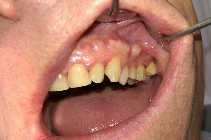
Buccal exostosis occurs on the buccal, or cheek side, of the mouth. Bony exostoses are not usually present until the late teens and early adult years in approximately 3% of the adult population, occurring more commonly in females. Many bony exostoses will continue to grow slowly throughout life.4
While sometimes a nuisance, these bony protuberances do not pose any threat. In cases where the bony projections are larger, certain tasks, such as performing oral hygiene, may become harder. Often, eating can become uncomfortable, especially regarding harder foods or those with sharper edges.3 Intraoral radiographs at the dental office will also present challenges and may be painful or sometimes next to impossible to obtain due to their interference with sensor placement.
Removal Methods
Tori or exostoses removal is not recommended unless it is clinically necessary. Removal becomes essential when they interfere with diet, speech, prosthodontics, and overall life satisfaction. This outpatient procedure can be performed using various methods, usually by an oral surgeon. Most methods can be traumatizing and painful, which often leads to patients postponing or completely avoiding treatment.5
Pre-operative planning with 3D models and molds may be necessary to help determine what method of surgical intervention is best suited for each individual.6
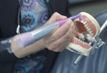
In 1988, Tommaso Vercellotti invented piezosurgery, a bone-cutting procedure that spares the soft tissue. This technique operates on ultrasonic micro-vibrations and pressure electrification. Piezosurgery is reported to create less discomfort for the patient, increase optimal visibility in the surgical field, and decrease blood loss compared to other methods.7
Another common method is using rotary instruments to remove excess bone. A soft tissue reflection is made to expose the exostoses fully, and a surgical carbide bur is used to score the superior aspects of the bone. Afterward, a monoplane chisel is used to remove the excess bone. Any sharp edges are smoothed prior to closing the surgical incision.4
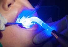
Conventional laser surgery has also been utilized for removal. After the bony overgrowth is exposed, a laser is used to excise the overgrowth. This method is less traumatic for the patient but does take longer to complete. Laser surgery also reduces trauma to surrounding tissues and increases comfort during the healing process.8
In 2002, a laser called the Er,Cr:YSGG was developed and approved by the FDA. This laser uses water and laser energy conjunctionally to excise hard tissue. This particular laser leaves behind little to no necrosis or tissue damage as it precisely cuts osseous tissue with no detrimental thermal transfer or charring at the gingival margin.5
The Dental Hygienist’s Role
A dental hygienist can visually identify tori and exostosis during an intraoral examination. Additionally, the bony protuberances will present on radiographs as well-defined round or oval over calcified areas, superimposing the roots of the teeth.2
Dental hygienists should note the appearance, location, and measurements of tori and exostoses in the patient’s chart. Dental professionals should bring these bony protuberances to the patient’s attention and provide any instruction on how to ensure the areas are cleaned around thoroughly to prevent inflammation.
If bony protuberances present as a solitary localized enlargement, benign and malignant bone pathologies, such as osteomyelitis, osteoma, and osteosarcoma, should be considered.1,3 Multiple bony growths not appearing in the classic tori and exostosis locations should be evaluated for Gardner syndrome. Other features of Gardner’s syndrome include multiple supernumerary teeth, cutaneous cysts or fibromas, or intestinal polyps, posing a high risk of malignancy.1-3
If a clinician has any doubt about the diagnosis of bony protuberances, a referral to an oral surgeon should be made.
In Closing
Dental professionals may take for granted that our patients understand or are aware of the oral anomalies that may be present. Even if the area presents no concern, bringing these bony protuberances to our patient’s attention is recommended. By doing this, we can extinguish any anxiety that may result when a patient stumbles upon these anomalies themselves. Furthermore, it validates that dental professionals have a wealth of knowledge about more than just teeth.
Before you leave, check out the Today’s RDH self-study CE courses. All courses are peer-reviewed and non-sponsored to focus solely on high-quality education. Click here now.
Listen to the Today’s RDH Dental Hygiene Podcast Below:
References
- Limongelli, L., Tempesta, A., Capodiferro, S., et al. Oral Maxillary Exostosis. Clinical Case Reports. 2019; 7(1): 222-223. https://doi.org/10.1002/ccr3.1918
- Medsinge, S.V., Kohad, R., Budhiraja, H., et al. Buccal Exostosis: A Rare Entity. J Int Oral Health. 2015; 7(5): 62-64. http://www.ncbi.nlm.nih.gov/pmc/articles/PMC4441241
- Mishra, I., Vijayalaxmi, N., Ramaswami, E., Jimit, D. Bony Exostoses: Case Series and Review of Literature. Acta Scientific Dental Sciences. 2018; 2(10): 64-67. https://actascientific.com/ASDS/pdf/ASDS-02-0328.pdf
- Exostoses and Tori. (n.d.). Exodontia Info. https://exodontia.info/exostoses-tori/
- Koceja, M.K. (n.d.). Atraumatic Excision and Ablation of Mandibular Tori Exostosis With Er,Cr:YSGG Laser Device. Live Dental. https://livedental.in/articles/general/285-atraumatic-excision-and-ablation-of-mandibular-tori-exostosis-with-ercrysgg-laser-device
- Bernaola-Paredes, W.E. Pereira, A.M., Albuquerque, Luiz, T.A., et al. An Atypical Presentation of Gigantiform Torus Palatinus: A Case Report. International Journal of Surgery Case Reports. 2020; 75: 66-70. https://www.ncbi.nlm.nih.gov/pmc/articles/PMC7490980/
- Pozzetti, E., Garibaldi, J., Cassinotto, E., et. al. Piezosurgery Removal of Mandibular Tori: A Case Series. Archives of Clinical and Medical Case Reports. 2023; 7: 290-294. https://www.fortunejournals.com/articles/piezosurgery-removal-of-mandibular-tori-a-case-series.html
- Rocca, J.P., Raybaud, H., Merigo, E., et al. Er:YAG Laser: A New Technical Approach to Remove Torus Palatinus and Torus Mandibularis. Case Reports in Dentistry. 2012; 2012: 487802. https://www.ncbi.nlm.nih.gov/pmc/articles/PMC3390044/


