Researchers have been utilizing tree rings for years to determine a tree’s age and the environmental factors that influence the development of a tree. In school, you may possibly recall learning this information yourself.
However, did you know human teeth also have “rings” similar to trees? Researchers are now concluding that these markings can be used to determine what types of stress a person may have endured and intervene by preemptively providing therapy to offset future physiological issues.
A Quick Review on Tooth Structure, Development, and Eruption
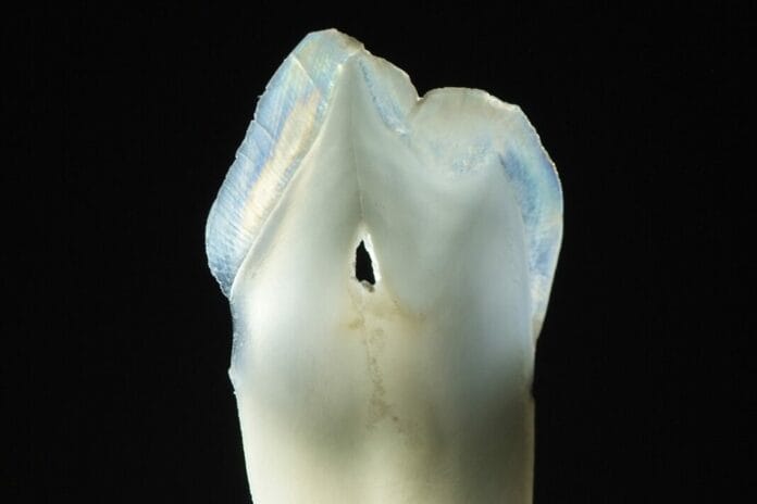
The human tooth is much like an ice cream sandwich and has several layers. Enamel, the outermost layer of the crown of a tooth, is the hardest, most highly mineralized tissue in the human body.1
The next layer that supports the structure of enamel is dentin. Unlike enamel, dentin exists in both the crown and roots of the tooth. Encased in the dentin are dentinal tubules that assist with “communication” for the surrounding tissues, such as the pulp where nerves lie.2 In the root of the tooth, the dentin is covered with a layer of cementum. Cementum is responsible for the attachment of the tooth to the bony socket.3
The pulp is the core of the tooth and contains not only the nerve but also blood vessels, dentin-forming cells, and odontoblasts.3
Humans develop two sets of teeth in their lifetime. The primary (baby) teeth began developing in the fetus at approximately six weeks gestation. The succedaneous or permanent teeth also began to develop early in gestation at approximately nine to 10 weeks.4 The hard tissue surrounding the tooth is formed at three to four months gestation.5
Teeth began erupting into the oral cavity as early as six months and until 33 months.5 Primary teeth begin exfoliation around age six through 12 on average. Permanent teeth typically erupt around six years of age, and the final root develops in the 3rd molars or “wisdom teeth” at ages 18 to 25.4,6
The Creation of Growth Marks
During the final stages of tooth development, dentin-producing cells called odontoblasts and enamel-producing cells called ameloblasts secrete proteins that create growth marks visible in the completed tooth crown. These markings are similar to the rings of a tree and act as permanent records within our teeth during the development process.3
Cross striations record daily growth, while long-period growth lines, called striae of Retzius, record weekly growth. These growth marks permanently record different phases of development because each tooth develops in a specific time window during ontogeny.3
Researchers can view daily and weekly development to piece together a continuous record of growth using a primary central incisor (with root intact) and a permanent second molar. A primary central incisor will represent development from prenatal life to age two postnatally, and permanent second molars will show development up to 14 to 16 years of age. Because natural exfoliation of the primary teeth does not include the root of the tooth, the timeline will be shortened when viewing these particular cases.7
Stress Lines in Teeth
When particular physical stressors occur, teeth development can be impacted. Common precursors are poor nutrition, disease, and ingested toxicants. These physiological stressors have been shown to produce growth marks known as stress lines.7
Enamel hypoplasia is one of the most commonly studied developmental defects.5 Enamel hypoplasia is a surface defect of the tooth crown that is caused by a disturbance of enamel matrix secretion, defective calcification, or defective maturation that appears as pitting, groves, or the complete absence of enamel.8
One of the most studied stress lines of the teeth is the neonatal line, which indicates an individual’s birth.5 Neonatal lines are produced due to the biological stress of a sudden change from intrauterine to extrauterine life. This represents the first formation of enamel after birth, separating prenatal enamel from postnatal enamel formation.9
This line is present in 90% of primary teeth in children and is used by anthropologists and forensic experts to determine causes and time of death in infants. Work by Andra and Smith revealed that using the neonatal line as a benchmark can determine other physical environmental factors such as injury, infections, extreme cold weather, and exposure to heavy metals and organic chemicals.7
Researchers are now studying how additional factors may impact teeth. Preliminary evidence suggested that psychosocial factors such as changes in family (divorce, death) and experiences of deprivation or threat (physical and sexual abuse, neglect, trauma) may present themselves as stress lines in the enamel.7
Using Stress Lines as Mental Health Risk Biomarkers
Studies on nonhuman primates revealed a connection between potential psychosocial stressor markers and disrupted tooth development. Nonhuman primates have two sets of teeth that develop incrementally and exhibit time-resolved growth marks, making these studies comparable to human teeth.7
Primates also undergo social stressors similar to those of humans. One study, in particular, evaluated captive juvenile rhesus macaques (monkeys) and showed a positive association between enamel stress lines and social stressors such as being separated from their mothers.7
A study of 47 teeth from a skeletal collection of 15 people aged 25-69 with a well-known medical and life history revealed that telltale signs of life events were recorded in the teeth. This particular study focused on the cementum and found that different colored rings within the cementum corresponded to the ages at which individuals underwent significant life and biological events with high accuracy. Researchers conclude that systemic illnesses and events such as incarceration could also be detected.10
Researchers suggest that stress lines within enamel could be used as biomarkers to predict risk for later psychiatric disease of the individual the teeth belong to. TEETH (Teeth Encoding Experiences and Transforming Health) is a model that proposes that early-life stressors disrupt multiple development processes and that primary and permanent teeth may serve as dual markers of both past psychosocial stress exposure and future mental health risk.7
These markers could be used to predict the risks of various mental/behavioral disorders, including schizophrenia, autism spectrum disorder, and attention deficit/ hyperactive disorder. With analysis of teeth, early intervention could be used to treat adolescents.7
Researchers suggest taking advantage of three opportunities within the first two decades of life to study human teeth. It is proposed that dentists and doctors could use these opportunities to send collected teeth off to be analyzed. The three opportunities include:7
- When primary teeth are naturally shed, beginning at ages six to eight: This is prior to the onset of puberty, a known high-risk period for major depressive disorder (MDD).
- Primary and permanent teeth extracted for orthodontic treatment: This typically happens right before the age of 13, which is immediately prior to the onset of MDD.
- When 3rd molars are extracted, typically between the ages of 15 and 20: This is the developmental stage when approximately 25% of MDDs occur.
In Closing
The Dunn Lab is continuously studying stress markers on teeth under the direction of Dr. Erica Dunn. She hopes these studies will impact children’s health by detecting future health risks. She encourages parents to donate their children’s teeth to science here. Dental professionals can help share this information with parents/caregivers to help assist in the ongoing studies. Collectively, we can support researchers in developing answers with this potentially life-changing information.
Before you leave, check out the Today’s RDH self-study CE courses. All courses are peer-reviewed and non-sponsored to focus solely on pure education. Click here now.
Listen to the Today’s RDH Dental Hygiene Podcast Below:
References
- Beniash, E., Stifler, C.A., Sun, C.Y., et al. The Hidden Structure of Human Enamel. Nature Communications. 2019; 10(1): 4383. https://doi.org/10.1038/s41467-019-12185-7
- Ponce, E.H., Sahli, C.C., Vilar, J.A. Study of Dentinal Tubule Architecture of Permanent Upper Premolars: Evaluation by SEM. Australian Endodontic Journal. 2001; 27(2): 66-72. https://doi.org/10.1111/j.1747-4477.2001.tb00343.x
- Goldberg, M., Kulkarni, A.B., Young, M., Boskey, A. Dentin: Structure, Composition and Mineralization. Frontiers in Bioscience (Elite Edition). 2011; 3(2): 711-735. https://www.ncbi.nlm.nih.gov/pmc/articles/PMC3360947/
- Sheldahl, L. C. (2020). Tooth Development. In L. C. Sheldahl, Histology and Embryology for Dental Hygiene. Open Oregon Educational Resources. https://openoregon.pressbooks.pub/histologyandembryology/chapter/chapter-8-tooth-development/
- Anatomy and Development of the Mouth and Teeth. (n.d.). Johns Hopkins Medicine. https://www.hopkinsmedicine.org/health/wellness-and-prevention/anatomy-and-development-of-the-mouth-and-teeth
- Developmental Data for Primary and Secondary Teeth. (n.d.). Pocket Dentistry. https://pocketdentistry.com/developmental-data-for-primary-and-secondary-teeth/
- Davis, K.A., Mountain, R.V., Pickett, O.R., et al. Teeth as Potential New Tools to Measure Early-Life Adversity and Subsequent Mental Health Risk: An Interdisciplinary Review and Conceptual Model. Biological Psychiatry. 2020; 87(6): 502-513. https://www.ncbi.nlm.nih.gov/pmc/articles/PMC7822497/
- Kanchan, T., Machado, M., Rao, A., et al. Enamel Hypoplasia and Its Role in Identification of Individuals: A Review of Literature. Indian Journal of Dentistry. 2015; 6(2): 99-102. https://www.ncbi.nlm.nih.gov/pmc/articles/PMC4455163/
- Janardhanan, M., Umadethan, B., Biniraj, K., et al. Neonatal Line as a Linear Evidence of Live Birth: Estimation of Postnatal Survival of a New Born From Primary Tooth Germs. Journal of Forensic Dental Sciences. 2011; 3(1): 8-13. https://www.ncbi.nlm.nih.gov/pmc/articles/PMC3190441/
- Cerrito, P., Bailey, S., Hu, B., Bromage, T. Parturitions, Menopause and Other Physiological Stressors Are Recorded in Dental Cementum Microstructure. Scientific Reports. 2020: 10: 5381. https://www.nature.com/articles/s41598-020-62177-7

