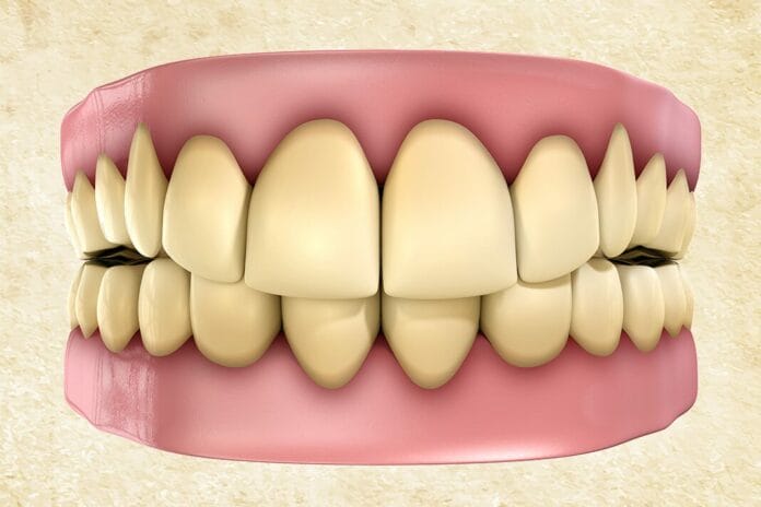
We have all seen firsthand that teeth become stained – some more than others. Our patients know and see it, too. They will let you know at their hygiene appointments and point out they do not like the color of their teeth.
There are many intrinsic and extrinsic causes for tooth discoloration, so identifying the etiology can help determine whether an appropriate intervention is indicated. If indicated, the etiology can also provide direction for treatment options and patient recommendations for prevention.1,2
As a quick refresher, intrinsic stains occur from changes in the structural composition or thickness of enamel. Endogenous intrinsic causes include tooth trauma (injury), disturbances in tooth development, fluorosis, or can be drug-induced.1,2
Restorative materials, endodontic treatment, and dentinal stains (i.e., secondary dentin formation, arrested decay, or active decay) are examples of exogenous causes. Enamel attrition and erosion resulting in thinner enamel can allow the yellow hue of dentin to show through, so teeth may appear more yellow or gray and are also categorized as exogenous.1,2
Extrinsic stain occurs on the external surface of the tooth. Direct extrinsic stains are caused by compounds or organic chromogens that attach to the acquired pellicle and cause discoloration. Extrinsic stains that result from a chemical interaction with the tooth surface are categorized as indirect.1,2
Yellow, green, black line, brown, and tobacco extrinsic stains are the most frequently observed. Less common are orange, red, and metallic stains.2
Below are eight common causes of extrinsic stains that may be seen in hygiene patients.
1) Poor homecare
We have seen firsthand how one’s home care and biofilm control can cause staining in the oral cavity. Sometimes, biofilm that has been missed several times can also be stained yellow, green, brown, and, in rarer cases, orange and red. Stains associated with poor oral hygiene are generally considered direct extrinsic stains.2
Yellow stain associated with poor oral hygiene is more common in older adults and is often associated with dietary choices and/or tobacco use.2
The distribution of stains associated with poor oral hygiene can range from localized to generalized. It is likely dependent on how poor the individual’s hygiene is, as calculus and biofilm can retain stains. The more present, the more generalized the stain may appear.2
2) Chromogenic bacteria (black line stain)
Chromogenic bacteria can cause staining. It can present in multiple shades, including green, dark brown, and black. In rarer cases, it can appear red or orange.2
The exact mechanism of how chromogenic bacteria cause staining is not well understood. It is proposed that hydrogen sulfide produced by the bacteria interacts with iron in the saliva, which leads to staining.3
The distribution of stains due to chromogenic bacteria varies and includes:2
- Line along the gingival third near the gingival margin
- Facial gingival third of the maxillary anterior teeth
- Irregular coverage of flat tooth surfaces
- Streaked in lines of the enamel or grooves of posterior teeth
Stains from chromogenic bacteria can affect children as well as adults. It can accumulate on both permanent and primary teeth.2
3) Smoking
This may be an obvious one, but it is a great opportunity for patient education regarding oral health risks. Not only can cigarettes and cigars cause staining, but vaping and cannabis can also contribute to extrinsic staining.2,4
Tobacco stain appears light brown to leathery brown or black. Considered a direct extrinsic stain, it is incorporated in biofilm and calculus. Additionally, heavy tobacco stain, particularly from smokeless tobacco, may penetrate enamel irregularities and become exogenous intrinsic stains.2
The distribution of stain appears as a narrow to wide tar-like band that follows the contour of the gingival margin. The band is not at the gingival margin, just slightly above it. It occurs primarily on the lingual surfaces and can cover the cervical third and extend to the central third of the crown of the tooth.2
Vaping can contribute to extrinsic stains due to the release of metal ions, pigments, and particles when the e-liquid is heated. Studies have shown little difference in extrinsic stains when traditional cigarettes were compared to e-cigarettes.4
Smoking cannabis can cause greenish staining. Often, staining occurs due to the acquired pellicle or dental biofilm becoming stained.2
Other predisposing factors include poor oral hygiene and the presence of calculus and biofilm, which facilitates stain adherence and can increase staining.2
4) Fruits and vegetables
Of course, the good, healthy foods! Have you seen a patient with tons of stains but they do not drink coffee or tea or smoke? I had a patient years ago with a mystery stain with no obvious origin. The next question I asked was if they consumed a lot of fruits or vegetables or drank smoothies. We discovered it was a protein shake with dark leafy vegetables, which the patient had recently started since her last recall appointment.
It has been suggested that an increased prevalence of black line stain can be seen with the consumption of iron-rich fruits and vegetables.3 Examples of iron-rich vegetables include broccoli, string beans, cabbage, brussels sprouts, and dark leafy greens such as spinach, collard, kale, and dandelion.5
Examples of iron-rich fruits include prunes and prune juice, dates, figs, and raisins.5
The dark chromogenic pigmentation of vegetables like beets or berries such as blueberries, blackberries, and pomegranates can cause stain. Cranberry, grape, and other dark-pigmented fruit juices can also cause staining.6 Besides chromogenic pigmentation, many berries also contain tannins (a polyphenol), which can result in tooth staining.1,7
5) Beverages
When it comes to beverages staining teeth, the culprit is usually the tannins found in the beverage. Coffee, red wine, and teas all contain tannins, which are the major contributing factors to tooth stains caused by these beverages.2
Interestingly, the stain caused by the tannins in different beverages may appear in different shades. For instance, red wine causes a gray stain, black tea and coffee stain yellow or brown, while green tea can cause either green or yellow staining.2,6,8 Fruit juices with dark pigment can cause a purplish stain.2
Acidic beverages can contribute to staining by increasing the roughness of enamel (decalcification/erosion). This facilitates easier deposition of chromogenic agents from pigmented beverages, leaving the enamel more prone to staining. For example, staining from colored carbonated beverages (i.e., Coca-Cola, Pepsi) is mainly due to phosphoric acid, which decalcifies, and chromogens, which act on the etched enamel, causing a brown discoloration.9
6) Antimicrobial agents
Some antimicrobial agents can cause extrinsic stains, such as chlorhexidine, cetylpyridinium chloride (CPC), copper salts, potassium permanganate, and stannous fluoride.
A dental hygienist’s worst nightmare is chlorhexidine stain. Chlorhexidine staining appears brownish on the tongue and tooth surfaces, with staining more pronounced on proximal surfaces and surfaces less accessible due to poor biofilm management. Exposed root surfaces tend to form stains from chlorhexidine more rapidly.2 Certain dietary choices like coffee, tea, and wine may worsen chlorhexidine staining.2,9
Chlorhexidine itself does not cause stain but instead binds dietary chromogens to teeth and mucosal surfaces, causing a colored layer. Likewise, staining can occur from the long-term use of mouthrinse that contains CPC.9,10
Additionally, mouthrinses that contain copper salts or potassium permanganate can cause staining. Mouthrinses with copper salts cause a green stain, while those with potassium permanganate cause a violet-black or brown stain. The mechanism that causes the staining is believed to be the interaction between the metallic compounds and dental biofilm.11
Toothpaste and mouthrinses that contain stannous fluoride can cause extrinsic stains. Staining occurs due to the improper and incomplete stabilization of stannous ions.12 The stain appears light brown, sometimes yellowish. The stain forms on the pellicle due to a chemical reaction between the tin ions in the stannous fluoride and chromogens and pigments in the diet.2,13
7. Swimming
Frequent swimming in pools treated with chlorine or bromine can cause yellowish or dark-brown stains on the facial surfaces of the maxillary and mandibular anterior teeth.2
The stain occurs due to the chlorine in the water breaking down salivary proteins, creating organic deposits on the teeth. The staining occurs on the acquired pellicle. Interestingly, though this stain can be removed by dental professionals, proper daily oral hygiene does not prevent the staining from occurring.14
8) Medications and supplements
Systemic medications and supplements can be a cause of tooth discoloration. Antimicrobial agents such as minocycline and doxycycline are thought to bind to the glycoprotein of the pellicle, causing extrinsic staining. This occurs more frequently in individuals with poor oral hygiene. These medications have also been indicated in intrinsic stain as well.11
Additionally, amoxicillin-clavulanic and linezolid have been linked to extrinsic “pseudo-discoloration.” Pseudo-discoloration refers to the altered oral microbiome that occurs when certain antimicrobial agents are taken. In some cases, the antimicrobial agent allows an overgrowth of chromogenic bacteria, which causes extrinsic staining.11
Iron supplements, specifically in liquid form as iron salts, can cause staining. The stain appears greenish-black or brown-black in color. Much like the reaction of copper salts in mouthrinses, a chemical reaction causes staining from iron supplements. The higher the contact surface of the tooth with the iron supplement, the greater the discoloration.15,16
Liquid chlorophyll is another supplement with the potential to cause staining. Chlorophyll supplements contain chlorophyllin rather than chlorophyll, which contains copper. As previously mentioned, copper can cause green staining due to the interaction of the metallic compounds and biofilm. The dark pigment of the liquid could also contribute to green staining.11,16,17
Management of Extrinsic Stain
The management of stain is dependent upon the causative factor. In some cases, simply improving oral hygiene can do the trick. Direct stains incorporated into the acquired pellicle can often be removed with proper brushing and interdental cleaning.2
Going back to the basics with proper home care and educating on proper brushing techniques and interdental cleaning may help reduce biofilm and, in turn, staining. It may be helpful to show patients the specific areas with stain and biofilm accumulation missed during daily home care. Recommending an electric toothbrush and finding interdental aids that work best for the patient based on their needs may also be helpful.2
The removal of indirect stains often requires intervention by dental professionals. Proper oral hygiene may be able to remove some stains, but in most cases, it cannot be removed in its entirety. Professional removal can often be achieved through scaling and/or polishing with an appropriate grit prophy paste.2
However, when stains are tenacious, it may be necessary to employ other techniques to avoid damage to tooth structure from abrasive polishing paste.2
Stain that is present on delicate root surfaces needs to be removed using the least abrasive technique possible. Prophy pastes may be too abrasive, and hand scaling may be too aggressive in these areas. Therefore, considering alternatives for these areas is imperative.18
Ultrasonic and sonic scalers have advantages for tough-to-remove stains on delicate tooth surfaces. Ultrasonic and sonic scaling are both efficient and effective for stain removal while reducing the loss of tooth structure compared to hand scaling and abrasive prophy pastes.18
Air polishing is another option for stain removal. Air polishing removes stains three times as fast as hand scaling with less fatigue. However, there are a few contraindications for air polishing, including those sensitive to sodium, those with respiratory problems, and those who wear contact lenses. In some cases, there are ways to mitigate the risks to these patients, including choosing a powder that is not sodium-based and offering safety goggles to those who wear contact lenses.18
In Closing
The shade of teeth color can be a concern for patients. Whitening is not always the right solution. Preventing extrinsic stains could require changes or modifications to a patient’s home care, diet, or lifestyle. More frequent recalls can be recommended as well.
Hygienists can evaluate changes in tooth color during intraoral exams and then be descriptive in our documentation. Asking the patient follow-up questions can help determine the cause of extrinsic staining and, in turn, guide prevention strategies and treatment options.
Before you leave, check out the Today’s RDH self-study CE courses. All courses are peer-reviewed and non-sponsored to focus solely on high-quality education. Click here now.
Listen to the Today’s RDH Dental Hygiene Podcast Below:
References
- Sruthy Prathap, H., Rajesh, V.A.B, Rao, A.S. Extrinsic Stains and Management: A New Insight. J Acad Indus Res. 2013; 1(8): 435-442. https://www.researchgate.net/publication/265169002_Extrinsic_stains_and_management_A_new_insight
- McConnell, C. A., & Boyd, L. D. (2023). Dental Soft Deposits, Biofilm, Calculus, and Stain. In L. D. Boyd & L. F. Mallonee (Eds.), Wilkins’ Clinical Practice of the Dental Hygienist (14th ed., pp. 301-325). Jones & Bartlett Learning.
- Janjua, U., Bahia, G., Barry, S. Black Staining: An Overview for the General Dental Practitioner. British Dental Journal. 2022; 232(12): 857-860. https://www.nature.com/articles/s41415-022-4345-0
- Vohra, F., Andejani, A.F., Alamri, O., et al. Influence of Electronic Nicotine Delivery Systems (ENDS) in Comparison to Conventional Cigarette on Color Stability of Dental Restorative Materials. Pak J Med Sci. 2020; 36(5): 993-998. https://pmc.ncbi.nlm.nih.gov/articles/PMC7372672/
- 52 Foods High in Iron. (2023, March 15). Cleveland Clinic. https://health.clevelandclinic.org/how-to-add-more-iron-to-your-diet
- Lindberg, S. (2020, December 11). 9 Foods and Drinks That Can Stain Your Teeth. Healthline. https://www.healthline.com/health/foods-that-stain-teeth
- 19. Petre, A. (2023, October 23). What Are Polyphenols? Types, Benefits, and Food Sources. Healthline. https://www.healthline.com/nutrition/polyphenols
- Duron, A. (2020, October 1). Which Foods Actually Stain Your Teeth (and Which Don’t). Greatist. https://greatist.com/grow/foods-that-stain-teeth
- Sarembe, S., Kiesow, A., Pratten, J., Webster, C. The Impact on Dental Staining Caused by Beverages in Combination With Chlorhexidine Digluconate. European Journal of Dentistry. 2022; 16(4): 911-918. https://www.ncbi.nlm.nih.gov/pmc/articles/PMC9683888/
- Addy, M., Mahdavi, S.A., Loyn, T. Dietary Staining In Vitro by Mouthrinses as a Comparative Measure of Antiseptic Activity and Predictor of Staining In Vivo. J Dent. 1995; 23(2): 95-99. https://pubmed.ncbi.nlm.nih.gov/7738271/
- Thomas, M.S., Denny, C. Medication-Related Tooth Discoloration: A Review. Dental Update. 2024; 41(5): 440-447. https://www.dental-update.co.uk/content/clinical-dentistry/medication-related-tooth-discoloration-a-review/
- Li, Y., Suprono, M., Mateo, LR., et al. Solving the Problem With Stannous Fluoride: Extrinsic Stain. J Am Dent Assoc. 2019; 150(4S): S38-S46. https://pubmed.ncbi.nlm.nih.gov/30797258/
- Wade, W., Addy, M., Hughes, J., et al. Studies on Stannous Fluoride Toothpaste and Gel (1). Antimicrobial Properties and Staining Potential In Vitro. J Clin Periodontol. 1997; 24(2): 81-85. https://pubmed.ncbi.nlm.nih.gov/9062853/
- Moore, A.B., Calleros, C., Aboytes, D.B., Myers, O.B. An Assessment of Chlorine Stain and Collegiate Swimmers. Can J Dent Hyg. 2019; 53(3): 166-171. https://pmc.ncbi.nlm.nih.gov/articles/PMC7533807/
- Nazemisalman, B., Mohseni, M., Darvish, S., et al. Effects of Iron Salts on Demineralization and Discoloration of Primary Incisor Enamel Subjected to Artificial Cariogenic Challenge versus Saline Immersion. Healthcare (Basel). 2023; 11(4): 569. https://pmc.ncbi.nlm.nih.gov/articles/PMC9957417/
- Teoh, L., Moses, G., McCullough, M.J. A Review and Guide to Drug-Associated Oral Adverse Effects – Dental, Salivary and Neurosensory Reactions. Part 1. J Oral Pathol Med. 2019; 48(7): 626-636. https://pubmed.ncbi.nlm.nih.gov/31192500/
- Bowman, J., Seladi-Schulman, J. (2024, July 25). Benefits of Chlorophyll. Healthline. https://www.healthline.com/health/liquid-chlorophyll-benefits-risks
- Pieren, J. A. & Bowen, D. M. (2020). Tooth Polishing and Whitening. In J.A. Pieren & D. M. Bowen (Eds.), Darby and Walsh Dental Hygiene (5th ed., pp. 505-518). Elsevier.


