Acids can be introduced to the mouth from the beverages or foods a person eats, from gastric juices due to GERD or bulimia, and bacterial metabolites, among several other ways. Dental professionals are well aware of the effects of acid in the oral cavity and the importance of maintaining a neutral pH in the mouth.
As a quick review, acids can demineralize tooth structure, leading to dentinal hypersensitivity and dental caries. Research estimates that 25-30% of the adult population reports experiencing dentinal hypersensitivity. The World Health Organization recognizes untreated dental caries as the most common health condition globally. Further, virulent bacteria thrive in an acidic environment raising the risk for periodontal disease. According to the NIH National Institute of Dental and Craniofacial Research, 42.2% of adults over the age of 30 in the United States have periodontitis.
The large percentage of the population who suffer from untreated dental caries has researchers looking into new ways to measure the pH of biofilm, understand acid’s effects on tooth structure, and recreate tooth structure altogether.
Intelligent Detection of pH Levels
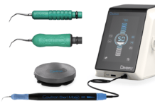
Researchers at the University of Washington are studying a prototype optical device that measures dental biofilm pH. The innovative technique entails the application of an FDA-approved chemical dye to the teeth. The optical device then emits LED light that measures the dye’s reaction with the light. It produces a pH reading that shows the pH level on a specific tooth and/or area of a tooth.
The measure helps dental professionals provide better treatment by knowing which area is most at risk for caries.
The research involved a population of a median age of 15, focusing more on pediatric patients whose enamel is thinner for better early warnings of demineralization.
Understanding Acid’s Activity Using 4D
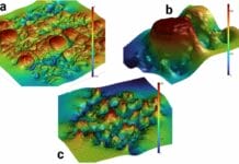
Little is understood about how acid damages dentin (intertubular dentin and peritubular dentin) and the differing rates of destruction at a microstructural level. To solve this, researchers at the University of Birmingham and the University of Surrey in the U.K. developed a new technique called “in situ synchrotron X-ray microtomography.”
The technique uses electrons to generate X-rays that scan dentin. It produced sub-micrometer resolution 3D images (a micrometer being one-thousandth of a meter, making the images very detailed) of dentin’s internal structure, highlighting the structural acid-induced changes over 6 hours. The images’ clarity has earned the study the name “4D studies.”
The goal of this technology and its findings is to understand further how acids demineralize different structures of dentin at different rates. It also looks to identify new ways to protect dental tissues and lead to new treatments.
Treatment Using Biological Enamel
Decayed tooth enamel is treated with restoration material because once past an incipient stage where remineralization is still possible, enamel cannot regenerate. Researchers are looking into recreating biological enamel to restore teeth instead of using synthetic restoration materials as we do currently.
In this technique, stem cells were taken from unerupted third molars and were used to create new stem cells. This research looked at recreating ameloblasts, the cells responsible for enamel production. However, other structures of the tooth and eventually the entire tooth might be able to be recreated using this study model.
This research provides a natural restoration instead of a less biocompatible synthetic restoration. Additionally, it alleviates possible recurrences like tooth necrosis, usually caused by synthetic treatments.
Before you leave, check out the Today’s RDH self-study CE courses. All courses are peer-reviewed and non-sponsored to focus solely on high-quality education. Click here now.


