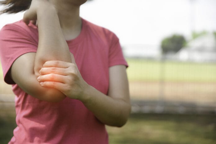
Both lateral and medial epicondylitis are afflictions that fall under the umbrella of musculoskeletal disorders (MSD). An MSD can be defined as “injuries affecting movement of the musculoskeletal system, which includes muscles, tendons, ligaments, nerves, discs, and blood vessels.”1
Lateral Epicondylitis
Lateral epicondylitis (LE) is caused by overuse and damage to both the extensor carpi radialis brevis (ECRB) muscle and tendon, which are located on the forearm. This specific duo of muscle and tendon helps keep the wrist and elbow balanced, especially when the forearm is held straight. As the elbow bends and straightens, the ECRB muscle and attaching tendon can rub against the lateral epicondyle or rather, the elbow, which can result in microtears in both the tendon and muscle.2-4
This condition is commonly referred to as “tennis elbow” because tennis players are at high risk for painful injury due to the position they frequently hold their forearm while striking with their racquets.2-4 It is important to understand that this can occur to anyone who may overuse this tendon and muscle group in their day-to-day life, such as dental hygienists.
Picture yourself, arm and wrist bent at a 90-degree angle, retracting the tongue while trying to gain access to the distolingual of #18 (possibly even the distolingual of #17). This puts you in the prime position to suffer from lateral epicondylitis.
Symptoms ─ Lateral epicondylitis can cause a wide range of symptoms that can include pain as well as a weakness in grip strength.2 This pain and weakness can occur while at rest but often become worse when the forearm is used excessively. A person suffering from LE may experience difficulty in everyday tasks such as holding items in their hands and turning a key or door handle.4
Diagnosis ─ In many cases, LE can be diagnosed based upon clinical presentation. The patient will undergo many questions that may include what their day-to-day activities and hobbies are, occupation, type of pain/symptoms being experienced, as well as information regarding the onset of symptoms (how long ago they started, is the pain/numbness/weakness occurring daily, do certain activities aggravate it, make it more comfortable, etc.).4
The physician may also utilize radiographs, MRIs, ultrasound, and electromyography (EMG) to make a diagnosis of LE. An x-ray can detect and remove arthritis from the potential diagnosis, while an MRI can provide a more detailed image and determine the injury’s extent. Ultrasound imaging can reveal changes or tears in the surrounding tendons as well as bone abnormalities. An EMG can be used to determine if any of the nerves surrounding the elbow are being compressed as a result of the disorder.2,4,5
Treatment ─ The goal of lateral epicondylitis treatment is to help with grip strength, restoring the area to pre-injury motion and stability, minimizing pain, as well as hopefully future injury prevention. As with most injuries, rest is the first step recommended.5
Due to the busy and nonstop nature of a hygienist’s demanding schedule, rest can prove to be impossible during the workday. So resting outside of the clinical setting may be the only option.
Other treatment modalities can include drug therapy with nonsteroidal anti-inflammatories, as well as steroid injections, ultrasound therapy, physical therapy, wearing a brace outside of work (prescribed by a doctor/physical therapist), electroshock therapy, and platelet-rich plasma injections (PRP).
PRP is a procedure where blood is withdrawn from the arm and is then placed into a centrifuge to allow platelets to be extracted. The platelets are then injected into the injured area and hopefully speeds up the healing process.2,4,5
If the above treatment modalities do not prove effective, surgery may be indicated. Depending upon the extent of the damage, LE may be treated surgically via either arthroscopic or open methods. Each patient’s recovery timeline will differ, but generally, they can return to pre-surgery activity four to six months after surgery.2 It is important to note that while LE surgical treatment can be extremely successful, some loss of strength may still be present post-surgery.2
Each patient’s degree of injury will vary, so their doctor will decide the best course of treatment, as well as the proposed healing timeline. This applies to hygienists as well.
Hopefully the above treatment results in a good outcome that allows for the hygienist to continue practicing. If not, looking into different career options may have to be considered if clinical dental hygiene is no longer an option.
Medial Epicondylitis
Medial epicondylitis is also considered an MSD. While LE affects the outside of the elbow, medial epicondylitis targets the inside of the joint. The medial common flexor tendon of the elbow is what is affected by this MSD.3 The tendon is attached to the “bump” that can be palpated along the inside of the elbow.
In these cases, patients complain of pain that radiates from the inside of the elbow all the way down to the wrist.6,7 This condition is commonly referred to as “golfer’s elbow” because it affects many golfers due to the force they use when they swing a golf club.7
I think most hygienists can agree that we don’t use the exact amount of force on our patients that it takes to swing a club. But we do use a fair amount of strength in this area, which puts us in danger of injuring the same tendon.
Symptoms ─ One of the most common symptoms in medical epicondylitis is soreness and discomfort along the inside of the elbow and forearm. This may be exacerbated when the patient attempts to curl their hand into a fist. A sense of weakness in the hands and wrists is another common complaint. A tingling sensation may also be felt from the elbow down into the ring and pinky fingers.5
Diagnosis ─ Often, a physical examination is all that is needed to diagnose medial epicondylitis. Similar to the diagnostic procedure used with lateral epicondylitis, questions regarding the patient’s day-to-day life and activities are important (What their profession is, what type of sports and activities they regularly engage in, etc.). Inquiries about when the symptoms started and what makes them worse or better are also important.
When a physical examination is not enough, other diagnostic tools that may be used include both radiographic and MRI imaging.4-7
Treatment ─ Recommendations for treatment of medial epicondylitis can be very similar to what is used to treat LE.
As mentioned above, the first step in treatment recommended is usually rest. Resting the affected area as much as possible can help alleviate symptoms and prevent further damage. Again, this can prove to be elusive to hygienists, so they will have to make time to rest whenever they can. Pain (typically over the counter) and anti-inflammatory medications, physical and or occupational therapy, ice, and PRP can be very successful in treating medial epicondylitis.4-7
If symptoms do not resolve with the above-mentioned treatment, surgery can be indicated.
A newer surgical procedure called TENEX utilizes an ultrasound-guided surgical technique to remove scar tissue. This type of surgery is considered minimally invasive.5
Why Dental Hygienists Should Care
Whether from holding our suction, mirror, or probe for hours on end, all while trying to wrestle the wildly strong buccinator and mentalis muscles, hygienists are in the prime position for the above-mentioned muscles and tendons become taxed and overused. In fact, one article says that “lateral epicondylitis is one of the most common overuse syndromes in primary medical care.”5
The Mayo Clinic goes on to state that one of the direct causes of medial epicondylitis is “forceful, repetitive occupational movements.”6 If this doesn’t summarize a day in the life of a dental hygienist, I’m not sure what else does.
Hopefully, bringing awareness to different MSD will help hygienists know what to be on the lookout for and when to seek medical attention.
Now Check Out the Self-Study, Peer-Reviewed CE Courses from Today’s RDH!
Listen to the Today’s RDH Dental Hygiene Podcast Below:
References
- Brillant, M.G.S, Doucette, H.J., Harris, M.L., Sentner, S.M., Musculoskeletal Disorders among Dental Hygienists in Canada. Can J Dent Hyg.2020 Jun; 54(2): 61–67. Retrieved from https://www.ncbi.nlm.nih.gov/pmc/articles/PMC7668274/#R4
- Tennis Elbow (Lateral Epicondylitis). (2020, August). American Academy of Orthopedic Surgeons. Retrieved from https://orthoinfo.aaos.org/en/diseases–conditions/tennis-elbow-lateral-epicondylitis/
- Kaider, K., Kiel, J. Golfers Elbow. Treasure Island (FL):StatPearls Publishing; 2021 Jan. Retrieved from https://www.ncbi.nlm.nih.gov/books/NBK519000/
- Tennis Elbow. (2021, February 25). Mayo Clinic. Retrieved from https://www.mayoclinic.org/diseases-conditions/tennis-elbow/symptoms-causes/syc-20351987
- Ma, K., Wang, H. Management of Lateral Epicondylitis: A Narrative Literature Review. Pain Res Manag.2020; 2020. Retrieved from https://www.ncbi.nlm.nih.gov/pmc/articles/PMC7222600/pdf/PRM2020-6965381.pdf
- Golfer’s Elbow. (2020, October 10). Mayo Clinic. Retrieved from https://www.mayoclinic.org/diseases-conditions/golfers-elbow/symptoms-causes/syc-20372868
- Medial Epicondylitis (Golfer’s and Baseball Elbow). (n.d.). Johns Hopkins Medicine. Retrieved from https://www.hopkinsmedicine.org/health/conditions-and-diseases/medial-epicondylitis-golfers-and-baseball-elbow


