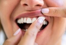
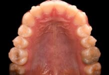
Black stain is “an external dental discoloration of bacterial origin, considered a special form of dental plaque.”1 The discoloration is characterized as a “dark line or an incomplete coalescence of dark dots localized on the cervical third of the tooth.”2
Black line stain can be difficult to manage for both patients and dental professionals alike. Many patients who experience black line stain require extra prophylaxis treatments to manage the stain.
The cause is somewhat elusive, as well as what exactly it is associated with regarding oral health and how to manage it.
Interaction with Iron?
Early published literature suggests that black line stain is an insoluble black ferric compound formed by the interaction between hydrogen sulfide produced by bacteria and iron.3 Additionally, previous theories postulated that chromogenic bacteria, such as P. gingivalis, contributed to the black pigmentation. However, recent studies show a “reduced microbial diversity in black stain as compared to standard plaque.”4
For many years, dental professionals relied solely on the idea that black line stain was associated with iron levels. In a recent study, the authors found that participants in the study presenting with black line stain had lower salivary and plaque iron levels than that of those without black line stain. Furthermore, this study found that the pH of the saliva was more alkaline than those that presented without black line stain. The study indicates that the pH of the oral environment likely plays a role in the presence of black line stain, and iron levels are less of an indicator.4
Other studies seem to confirm this finding, as it has been shown that individuals with black line stain have a lower risk of developing caries. In one study, the authors found a higher buffering capacity of saliva and a higher level of calcium in the saliva of patients presenting with black line stain.2 This is not a new concept; as far back as 1923, there was discussion of black line staining providing “immunity” to dental caries.5
What Type of Water?
Since these studies have found that a more alkaline oral environment contributes to the presence of black line stain, dental professionals should consider discussing what type of water the patient consumes. Alkaline water is all the rage, and as a dental professional, I understand the benefits of drinking water that is not acidic.
However, some of the bottled water companies tout a pH of 10, which could be a contributing factor to the development of black line stain. With that in mind, consider recommending water with a more neutral pH. Nestle Pure Life (pH 6.24) and Great Value bottled water (pH 6.04) were the most affordable brands I could find, with a pH relatively close to being neutral. Though this isn’t ideal, as a pH of 7 to 7.5 would be preferred, many bottled waters are much more acidic, which could cause more harm than good.6
If affordability is not a concern, FIJI (pH 7.57) and Evian (pH 7.59) are better choices.7,8 Just as we don’t want our patients to consume acidic water due to increased caries risk, we should steer them away from water that is too alkaline as well, especially if they are experiencing black line stain.
One study even recommended snacking between meals to reduce the pH level to a more neutral level. However, I feel that snacking is not a good habit to start or recommend as a dental professional. In extreme cases, recommending more acidic drinking water may be an option, but that, too, feels a little unethical to me as decay can lead to more serious dental problems, while black line stain is just not aesthetically pleasing.
Moreover, a study found that the oral microbiome of children with black line stain had a more balanced oral microbiome, better oral hygiene, and a lower prevalence of dental caries.9 Though black line stain may be frustrating for the patient and dental hygienists, these studies indicate there is a protective aspect against caries due to the oral environment.
The Metagenomics of Iron
To make things even more difficult to dissect, a study used metagenomics to characterize the composition of black line stain and found high levels of iron in the stain.1 A quick definition for those wondering what metagenomics is, it is “the study of the structure and function of entire nucleotide sequences isolated and analyzed from all the organisms (typically microbes) in a bulk sample.” It is used to identify specific communities of microorganisms.10
As mentioned previously, other studies found low levels of iron in the saliva. I want to mention that just because there are low levels of iron in saliva, that does not mean that black line stain doesn’t have higher levels of iron when compared to white plaque. There are pathways that have yet to be identified that might play a role in the distribution – higher levels of iron are found in the black line stain, while lower levels of iron are found in the saliva.
For example, iron is an important mineral/element in our body. Iron plays multiple roles, and everyone needs to have an ideal level to remain healthy. When you look at the “iron wheel, published by Trace Elements,” you will see that calcium is an antagonist of iron.11 Therefore, if there is a higher level of calcium in the saliva, which was reported in one study, it is reasonable to believe there would be a lower iron level in saliva. Since calcium inhibits iron absorption, it is plausible that salivary iron not being absorbed is being deposited on teeth in the form of black line stain.12
In no way am I implying iron isn’t being absorbed further in the GI tract and causing systemic concerns, as that is an entirely different mechanism. I’m simply highlighting that iron may remain on teeth at a higher level than in individuals with a lower level of calcium in the saliva.
As a full disclosure, this is strictly an opinion regarding the pathway by which this could occur. To my knowledge, there are no studies to support this theory. I wanted to clarify how it could be possible that these two studies that seem to contradict one another could both be accurate in their findings. Each study had a different endpoint leaving much to be discovered regarding black line stain etiology. In short, the etiology of black line stain is more complex than one might assume.
Dental Management of Black Line Stain
This leaves the most important question: What can we do for patients who present with black line stain? The three main factors that were highlighted in the studies associated with black line stain were:
- Consuming water with high iron content
- Consuming water that is too alkaline (pH above 7)
- Higher salivary pH associated with higher calcium levels in saliva
The best modifier would be to manage the amount of iron in their drinking water and/or encourage patients to consume more neutral water than alkaline. If your patients presenting with black line stain report drinking tap water, they may need to switch to bottled water to reduce the iron levels in their water or get a filter that will filter some of the iron out of the water. If your patients with black line stain report drinking alkaline water, encourage them to switch to tap water (assuming the water does not have high levels of iron) or bottled water with a neutral pH to reduce the risk of black line stain.
Patients and dental hygienists despise black line stain. Though it is not associated with poor oral health, disease onset, or disease progression, it is quite annoying aesthetically. Our best tool currently is interviewing patients with black line stain to determine the source of drinking water and making recommendations accordingly. Future studies may bring more options to light, but as it stands, the tools we have to manage black line stain are limited.
Before you leave, check out the Today’s RDH self-study CE courses. All courses are peer-reviewed and non-sponsored to focus solely on high-quality education. Click here now.
Listen to the Today’s RDH Dental Hygiene Podcast Below:
References
- Veses, V., González-Torres, P., Carbonetto, B., et al. Dental Black Plaque: Metagenomic Characterization and Comparative Analysis with White-plaque. Scientific Reports. 2020; 10(1): 15962. https://doi.org/10.1038/s41598-020-72460-2
- Żyła, T., Kawala, B., Antoszewska-Smith, J., Kawala, M. Black Stain and Dental Caries: A Review of the Literature. BioMed Research International. 2015; 2015: 469392. https://doi.org/10.1155/2015/469392
- Reid, J.S., Beeley, J.A., MacDonald, D.G. Investigations into Black Extrinsic Tooth Stain. Journal of Dental Research. 1977; 56(8): 895-899. https://doi.org/10.1177/00220345770560081001
- Ortiz-López, C.S., Veses, V., Garcia-Bautista, J.A., Jovani-Sancho, M.D.M. Risk Factors for the Presence of Dental Black Plaque. Scientific Reports. 2018; 8(1): 16752. https://doi.org/10.1038/s41598-018-35240-7
- Pickerill, H.P. A Sign of Immunity. British Dental Journal. 1923; 2: 967-968.
- Orr, T. (n.d.). Brands of Bottled Water that are Acidic. Water Purification Guide. https://waterpurificationguide.com/brands-of-bottled-water-that-are-acidic/
- Bottled Water Quality Report [Reference Report: #A00357257]. (2020, April). Fiji Water. https://www.fijiwater.ca/content/dam/fijiwater/faq/2020_CA_Water_Quality_Report_FINAL2_English.pdf
- Evian Natural Spring Water – Annual Water Quality Report. (2020, December 15). Evian. http://dev1.evian.com/wp-content/themes/evian/assets/report_files/evian_Bottle_Water_Quality_Report_2020.pdf
- Zhang, Y., Yu, R., Zhan, J.Y., et al. Epidemiological and Microbiome Characterization of Black Tooth Stain in Preschool Children. Frontiers in Pediatrics. 2022; 10: 751361. https://doi.org/10.3389/fped.2022.751361
- Metagenomics. (2024, January 8). National Human Genome Research Institute. https://www.genome.gov/genetics-glossary/Metagenomics
- Iron Wheels. (1988). Trace Elements. https://www.traceelements.com/docs/Iron%20Wheels.pdf
- Puri, S., Li, R., Ruszaj, D., Tati, S., Edgerton, M. Iron Binding Modulates Candidacidal Properties of Salivary Histatin 5. Journal of Dental Research. 2015; 94(1): 201-208. https://doi.org/10.1177/0022034514556709


