One of the most effective ways to gain case acceptance in the dental practice is through “co-planning” each dental patient’s treatment. Co-planning is essentially a broader concept of theoretical hand-holding and guiding your patient along as you work together to assess, identify, and plan the best course of care for specific oral health concerns.
In pediatrics, we often use the “tell-show-do” method. For example, we tell the child we have an electric toothbrush that is going to tickle their teeth. Then we show them our prophy angle and perhaps even let them feel it on top of their fingernail. At that point, we progress to using it on one of their teeth.
With adults, co-planning is essentially equivalent to show-tell-do for children ‒ but on a much higher scale. No one enjoys having something told or done to us that we don’t understand or relate to. When conditions such as periodontal disease or caries are present, we need not assume that the patient already understands the gravity of their position.
Putting Co-planning into Action
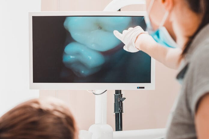
Here’s where intraoral cameras and enlarged radiographs come into play. By always ensuring that the imagery is within the eyesight of the patient, you can visually point out areas in their mouth that differ from those around them, such as bone levels, interproximal shadowing, or discolorations inside of occlusal pits. Even if they aren’t due for radiographs that day, taking photographs is extremely valuable.
First, talk about what the condition is and how it affects their mouth. But keep it simple. Now is not the time for a full-blown lesson on the etiology of a specific disease. Simple statements such as “When we see areas of bone loss, we want to specifically watch out for periodontal symptoms, since periodontitis is usually the leading cause of tooth loss.” Or “Any time we see shadows on X-rays, we want to rule out cavities since decay is known to spread quickly or even into adjacent teeth.”
Second, share the imagery with the patient. As you’re looking through each radiograph or photo, say what you see. “This looks healthy” is completely fine if there’s nothing alarming. But when you do notice something, pause and show them. You might say, “Hmm, see this shadow there? It looks like it could possibly be decay forming in the outer layer of your enamel. See how it looks compared to the tooth next to it? I’ll want to have the doctor check it during your exam.”
At this point, you’ve already guided the patient into co-diagnosis of their condition.
Finally, once the doctor has formally diagnosed any specific condition, identified what the treatment plan should be, and then left the room, it’s up to you to reinforce the formal diagnosis and verbally walk through co-planning with the patient. Back up to where you introduced the condition and what it causes, review what you both saw on the radiograph/photo together, and then reinforce how the proposed treatment will help to address this specific problem before it gets any worse.
Why it Matters
It sounds simple and self-explanatory but, as dental professionals, we often fall into the habit of routine. During a routine, we often leave out the necessity of allowing patients to personally diagnose and plan their treatment under the supervision of the dental team. When we empower patients with the simple information that they need to understand their conditions, they naturally become more accepting of their recommended treatment.
Patients want to know the “why.” Why do they need it? Why does it matter? And why is it being recommended so strongly?
When you put them in the position of “seeing is believing,” patients can naturally be more reciprocal of the care your dentist is advising. But we usually can’t make it happen without the images to go alongside the information. If you’re not already co-planning with radiographs and especially intraoral cameras, this one minor change can have a huge impact on your practice’s case acceptance numbers from day one.


