When a posterior maxillary tooth has been lost, a physiological process begins that can cause the alveolar ridge to resorb. This process can result in such a great amount of bone loss that the maxillary sinus begins to drop lower than it was prior to the tooth being lost.
One of the negative side effects of this can be that the lack of bone can prohibit a patient from being a candidate for a dental implant due to the lack of bone height.1 Sinus augmentation is a surgical procedure that aims to correct this and allow the patient to proceed with implant placement surgery.
What Is a Maxillary Sinus?
The largest of the paranasal sinuses, the maxillary sinus, is composed of four walls, the base of which constitutes the lateral wall of the nose and an apex that continues to the zygomatic process.2 It is housed inside the maxillary bone lateral to the nasal cavity.2
The maxillary sinuses play a role in the vibrations of a person’s voice, the contour of the face, and helping to keep inhaled air moist and warm. These sinuses develop at 16 weeks gestation and continue to grow as secondary teeth erupt.2
Anatomy and Blood Supply
In 1910, Arthur S. Underwood discovered an important piece of maxillary sinus anatomy named the septa. Septa can occur as a result of an abnormality a person was born with or can happen as the sinus floor expands once a tooth has been lost.3
The septa are of critical importance when planning for dental implants since the presence and location of the septa can interfere with the implant placement. It can also put a patient at a higher risk for sinus tearing during surgery.3
Vascularization of the maxillary sinus is achieved via many pathways, including the infraorbital artery, posterior lateral nasal artery, and posterior superior alveolar artery, all of which are maxillary artery branches.4 Being aware of the locations of these arteries is essential to avoid damage or hemorrhaging.4
Sinus Pneumatization
Pneumatization of the sinus is defined as a physiological process that causes the sinus to increase in size. Studies have shown that the pneumatization process is induced after a posterior maxillary tooth has been extracted and can cause the floor of the sinus to be joined with the alveolar bone.2
Methods and Techniques
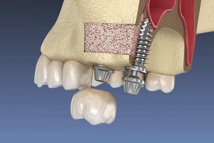
While the initial sinus augmentation surgery that originated in the 1970s differs from today’s procedures, the same general process is followed. During the sinus augmentation procedure, the floor of the sinus is lifted up, and bone grafting material is placed underneath the sinus to allow greater space for the dental implant to occupy.4,5
The two most common methods of sinus augmentation surgery used today include a lateral window technique, also known as a direct sinus lift, and an osteotome elevation, also known as an indirect sinus lift. The surgeon will determine which approach they use based on personal preferences as well as how much the sinus floor needs to be elevated.4,5
During the lateral window technique, a circular incision is made in the alveolar bone near the premolar/molar region using either hand or motorized instruments where the sinus membrane can be visualized entirely. The membrane is detached from the sinus wall, the sinus is lifted, and bone grafting material is placed underneath to elevate the sinus.4,6
During an osteotome elevation, a crestal incision is made, and a surgical flap is then opened, exposing the underlying alveolar bone. Then, a series of osteotomes are placed in the bone in order of smallest to largest via either a hammer or handpiece. As the osteotome sizes progress, the larger the incision becomes. Bone grafting material is placed under the sinus to push up and elevate the membrane.4,6
Depending on how much bone needs to be regenerated, a dental implant can be placed simultaneously during the sinus augmentation procedure, or the area may need to heal for four or more months before an implant can be placed.7
Risks and Complications
Perforation of the sinus membrane is the most common risk of sinus augmentation. This complication is estimated to happen between 7% to 35% of all sinus augmentation surgeries.4
The lateral window technique has a higher risk for sinus membrane perforation than the osteotome elevation technique because it requires a larger flap procedure and more precise surgical skill and capacity.5 Bleeding from either the sinus or the alveolar bone is another potential risk during these procedures.4
Movement of a dental implant into the maxillary sinus is also a potential complication post-sinus augmentation. It can occur any time after the surgery has been completed, including when the abutment is seated on the implant or even many years after implant placement. This can be caused by improper placement of the implant at the time of surgery, overuse of the drill during the procedure, or using more force than necessary.4 This is considered a dental emergency, and the implant must be removed immediately.4
This is why it is imperative that the surgeons performing these procedures be highly skilled, educated, use technology, and understand which patient population may have the greatest outcomes with the lowest possible complications.4
Contraindications for Sinus Augmentation
There are several contraindications for sinus augmentation procedures. Just like any other surgical procedure, some systemic conditions and habits may prevent a patient from being a qualified, healthy candidate. These can include a history of poor or delayed wound healing, uncontrolled diabetes, and heavy use of alcohol and tobacco.4
A few area-specific contraindications can include recurrent sinus infections, severe allergies, cancers of the sinus, other sinus surgical procedures, and previous radiation in the area.4
The Value of Cone-beam Computed Tomography
Dental professionals recognize the abundance of invaluable information that cone-beam computed tomography (CBCT) can provide. However, the technology is fundamental when it comes to evaluating the sinuses. “Cone-beam computed tomography (CBCT) provides more precise dimensions of the residual bone height and density. It also provides information about the maxillary sinus membrane, arterial passages in the lateral sinus wall, pathologies of the maxillary sinus, and the presence of septa.”4
The CBCT can not only identify the location and presence of septa but can also show their height, width, and orientation.4
In Closing
As dental hygienists, we know the importance of restoring patients’ posterior occlusion. Our patients have numerous reasons why treatment is delayed once a tooth has been lost. Unfortunately, this can result in the loss of adequate bone height, which can complicate placing a dental implant, and patients may have questions during hygiene treatment.
Luckily, we have predictable options for helping patients restore their smile, occlusion, and overall dental health.
Before you leave, check out the Today’s RDH self-study CE courses. All courses are peer-reviewed and non-sponsored to focus solely on pure education. Click here now.
Listen to the Today’s RDH Dental Hygiene Podcast Below:
References
- Haggag, M., Askar, O., Attia, A. Chronic Sinusitis: Does It Contraindicate Lateral Maxillary Sinus Lift? Egyptian Journal of Oral and Maxillofacial Surgery. 2022; 13(1): 1-8. https://omx.journals.ekb.eg/article_246682.html
- Alqahtani, S., Alsheraimi, A., Alshareef, A., et al. Maxillary Sinus Pneumatization Following Extractions in Riyadh, Saudi Arabia: A Cross-sectional Study. Cureus. 2020; 12(1): e6611. https://www.ncbi.nlm.nih.gov/pmc/articles/PMC6957056/
- Dandekeri, S.S., Hegde, C., Kavassery, P., et al. CBCT Study of Morphologic Variations of Maxillary Sinus Septa in Relevance to Sinus Augmentation Procedures. Ann Maxillofac Surg. 2020; 10(1): 51-56. https://www.ncbi.nlm.nih.gov/pmc/articles/PMC7433935/
- Bathla, S.C., Fry, R.R., Majumdar, K. Maxillary Sinus Augmentation. J Indian Soc Periodontol. 2018; 22(6): 468-473. https://www.ncbi.nlm.nih.gov/pmc/articles/PMC6305100/
- Wimalarathna, A. Indirect Sinus Lift: An Overview of Different Techniques. Biomed J Sci & Tech Res. 2021; 33(4). https://biomedres.us/pdfs/BJSTR.MS.ID.005447.pdf
- Thube, D.M., Devkar, N., Vibhute, A.R., et al. Management of Maxillary Posterior Inadequate Ridge Height for Prosthetic Rehabilitation. J Dent Allied Sci. 2018; 7(1): 27-33. https://jdas.in/article.asp?issn=2277-4696;year=2018;volume=7;issue=1;spage=27;epage=33;aulast=Thube;type=3
- Sinus Augmentation. (n.d.). American Academy of Periodontology. https://www.perio.org/for-patients/periodontal-treatments-and-procedures/dental-implant-procedures/sinus-augmentation/


